See the Full Issue
July 2024
When a local zoo veterinarian called to request a tooth extraction for a North American black bear, Jan Bellows, DVM, DAVDC, DABVP (Canine & Feline), and his team discovered the bear had a fractured right lower canine tooth, common in bears due to their powerful jaws and the demands of their natural foraging behaviors.
See the Full Issue
Our local zoo veterinarian called to request a tooth extraction for a 15-year-old, 221 kg North American black bear who suffered a crown/root fracture of the right lower canine tooth. The animal care team observed the bear exhibiting discomfort, including decreased appetite and a reluctance to chew on his favorite enrichment items. Upon closer examination, the veterinary staff discovered a fractured right lower canine tooth, common in bears due to their powerful jaws and the demands of their natural foraging behaviors.


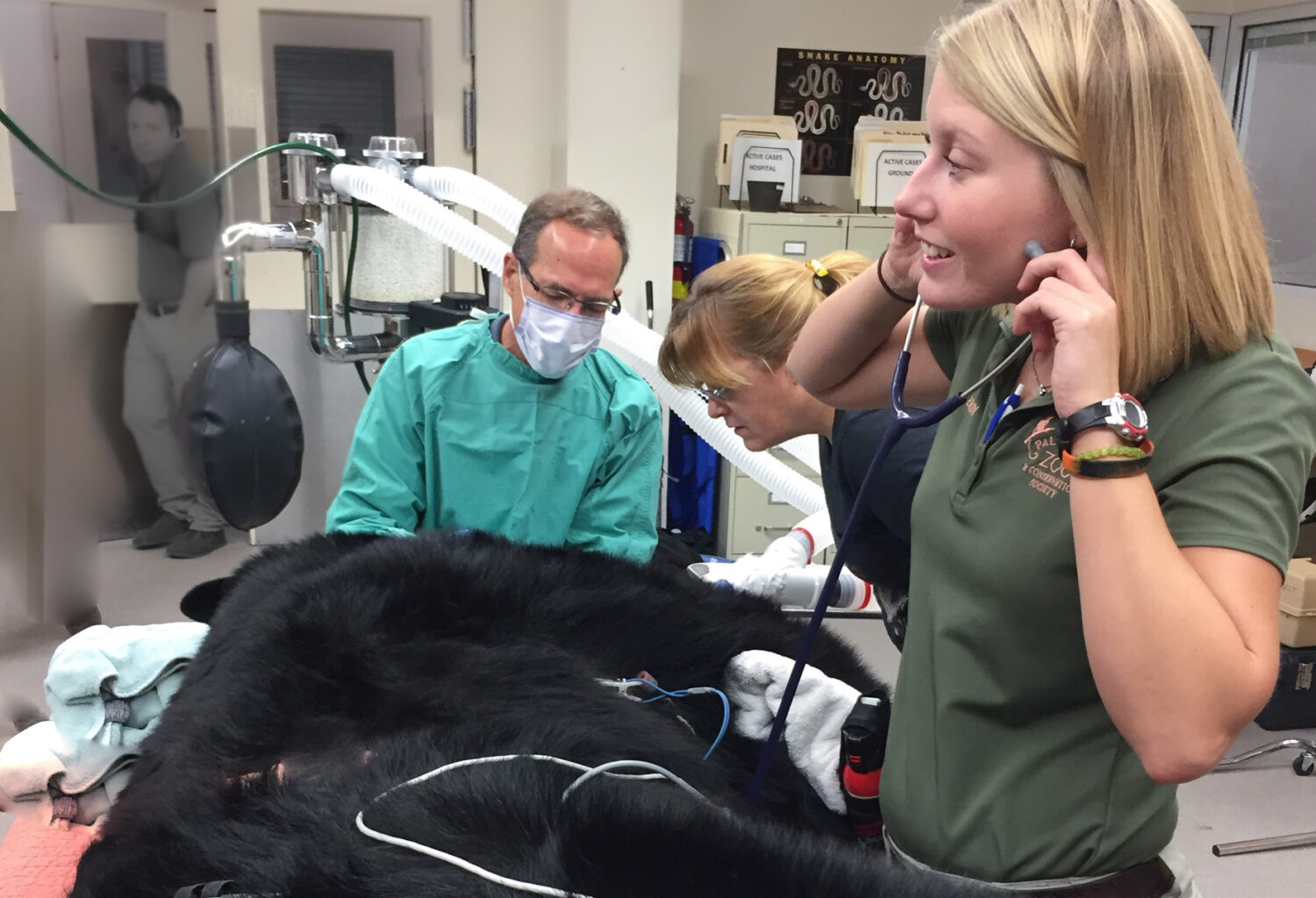
Once the zoo staff confirmed the bear was healthy enough for anesthesia, an intramuscular (IM) injection of medetomidine hydrochloride (0.03 mg/kg) was administered as a preanesthetic. Ten minutes later, tiletamine (4 mg/kg) was administered IM for induction. The bear was intubated, and anesthesia was maintained with isoflurane and oxygen (Figure 1).
Examination under anesthesia confirmed a chronic crown root fracture with a significant vertical crown segment still attached subgingivally (the slab). Once the vertical crown segment was removed, further examination could be conducted (Figure 2).
A middle mental nerve block with 4 mg/kg of bupivacaine hydrochloride (0.75%) was administered in addition to a splash block around the affected canine tooth to ensure the bear’s comfort and safety throughout the procedure. The fractured tooth slab was removed using a Molt#2 periosteal elevator designed to separate the fractured tooth segment from the surrounding tissues (Figure 3a and b). A 12 mm periodontal pocket and marked gingival enlargement were observed surrounding the mobile slab. The periodontal pocket was reduced to a 4 mm pocket after a gingivectomy was performed using a 15c scalpel blade.
An intraoral radiographic evaluation revealed minimal periapical lucency consistent with endodontic disease; it was determined that the fractured tooth was treatable through conventional root canal therapy with a good prognosis (Figure 4). A #3 barbed broach was inserted far into the pulp chamber and rotated to remove the tooth’s pulp (Figure 5, 6). The root canal and pulp chamber were disinfected with sodium hypochlorite to eliminate any remaining organic matter. Saline was then flushed into the canal to remove the chemical and ensure a clean and sterile environment. The root canal and pulp chambers were then carefully dried using paper points (Figure 7).
The cleaned canal was obturated (filled) with Gutta Flow 2® (Coltene), a biocompatible, flowable bactericidal dental material, followed by a gutta percha point to complete obturation (Figure 8a and b). A radiograph was exposed and examined, confirming an appropriate fill (Figure 9). Finally, the tooth was restored with a glass ionomer cement placed over the gutta-percha and light-cured flowable composite, a durable dental restorative material (Figure 10a and b).
The veterinary team closely monitored the bear following the successful root canal procedure to ensure a smooth recovery. Pain management medication (buprenorphine 0.02 mg/kg) was administered IM. Meloxicam (0.2 mg/kg) was administered orally once daily for five days. Over the next few weeks, the veterinary staff continued to assess his progress, closely monitoring the treated tooth for any signs of complications or infection.
This bear’s complicated crown/root fractured tooth was successfully saved; he returned to his routine, enjoying his favorite enrichment activities and maintaining a healthy appetite. His other dental abnormalities, including extraction of the right mandibular incisor and root canal therapy of the right maxillary canine, will be addressed when anesthetized for his upcoming annual wellness examination.
Photos courtesy of Jan Bellows