
See the Full Issue
April 2023
A new tool could provide easier diagnoses for lumps and growths
See the Full Issue
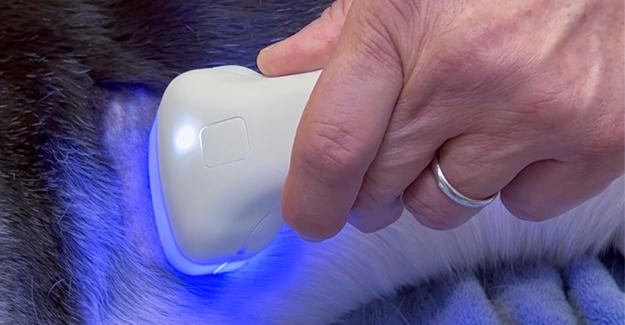
Photo 1 (Top): The blue light comes on during the heating phase, which only lasts a few moments.
Photo 2 (Bottom): A white light comes on during the scanning phase of the procedure.
I have encountered so many surprises when seemingly innocent looking lumps have gotten a low score and then turned out to be sarcomas, mast cell tumors, etc., that the number of surgeries I have performed has almost doubled. And these are the growths about which we may have said, “Let’s wait and see.”
In the US, there is an estimated 14.5 million skin growths on dogs detected and brought to the veterinarian’s attention every year. Yet only 40% of these undergo any kind of diagnosis. We can only venture a guess as to why, but I think there are several reasons we need to consider.
In the office of most practices, we only have the fine-needle biopsy (FNB) and our microscope to start the diagnostic process. This method has limitations in the type of diagnostic information we can get.
First, we are only getting a 1 mm wide sample out. Do we know if those cells are representative of the entire lump? Second, what is your confidence level of reading that slide? For me, anything beyond adipose tissue and the basophilic stippling that usually accompanies mast cell tumors falls outside my comfort zone. And lastly, if you find that you have just done a FNB on a cyst, you could be sending home an animal that is now leaking cystic material, or even worse, you have introduced bacteria into the cyst from the now open hole and you will see the dog back in a week with an infection you now have to explain and treat.
The next level of diagnosis is a biopsy punch. With this method, we are more likely to get a diagnosis because we are taking a several millimeter-wide swathe of the lump. We still face the issue of putting a hole in a cyst. And, a new issue arises: Biopsy punches can only go so far into tissue, so in the case of growths in obese dogs, growths that are surrounded by necrotic tissue, or that are just located deeper into the tissue, we may not get a sample that is representative of the lump. And of course, we need to—at the very least—sedate and provide a local anesthetic for pain management.
Photo 3: The HT Vista Device, screen on the left, scanning wand on the right
The most diagnostic procedure we have is to take the lump out and send it to a pathologist for histopathology. But then the question of margins arises before taking that lump out; how big should the margins be? If it is a malignancy and we don’t take both wide and deep margins, we will most likely be performing a follow-up surgery to get clean margins. A follow-up surgery is going to be an added expense and stress for the owner and more pain, fear, anxiety, and stress for the patient.
And if we decide to just do wide margins and it is a benign growth, we have created a longer, deeper and more painful incision that will also create an additional expense for the owner. And with this method of diagnosis, we now need to consider both the risk and adverse events depending on the location; those lumps that are on the face could result in disfigurement if a large enough area is removed, and those lumps on extremities may not have enough tissue to close the wound, even with advanced plastic surgery techniques like a z-plasty. And finally, there may be underlying structures such as major arteries and veins, or structures like salivary glands that you want to avoid.
The HT Vista is a noninvasive diagnostic device for dogs and cats that can screen a skin growth and make a determination if it is malignant or benign. It can look at growths that are on the surface of the skin, within the dermis, or that are subcutaneous. It cannot look at deeper growths: it can’t scan growths on internal organs, etc. It also cannot look at growths that are necrotic or have raw surfaces. But it can look at everything else.
Photo 4: Carefully shaved area prior to scanning
The science behind this device is based on how different tissues react to mild heating. The unique heat flow property is different in malignant growths than it is in either benign growths or normal skin tissue. Best of all, it has a 98% negative predictive value: this means that it was designed to always predict the malignancies and if it is not certain, it errs on the side of caution, asking the user to investigate further. This gives you complete confidence when it calls a growth benign.
The HT Vista must have an internet connection to connect with the algorithm and AI in order to make a diagnosis. The animal’s skin is prepped by clipping with a #40 blade: hair can interfere with heat absorption and dispersion giving a positive result when it is not. The handle (see photo 3) is then placed over all or a portion of the lump but including some normal skin for comparison purposes. A button is pressed, it gently heats the tissue for a few seconds, and then it scans the resulting diffusion and sends the findings off. This part takes about one minutes in addition to prepping the area for scanning. Within minutes you will get a result on a scale of 1–10, with 1–4 requiring further investigation and 5–10 benign.
Photo 5: The results are displayed on the screen of the device within minutes of finishing the scan
Mutzarella is a 12-year-old spayed female Border Collie mix who presented with a growth on her thorax near the axilla. The owner said the growth had been there for some time, but it had changed recently, becoming firmer and larger in size. The owner wanted it off, and we discussed using the HT Vista as a decision support tool for margin width and depth for surgical removal.
Step one was to shave as close as possible over the lump and some adjoining tissue (photo 4). Next, the scanner was placed over the lump and some adjacent normal tissue, the button was pushed, and a blue light came on for a few moments, showing that the tissue was being heated (photo 1). This was followed shortly after by a white light during the scanning phase (photo 2). Next, the machine asked us to highlight an area representing both the lump and normal tissue, and then we sent it off for analysis.
The answer came back with a score of 3, indicating probable malignancy (photo 5). Mutzarella was appropriately prepped and given a local block for a wide margin surgery.
There is nothing in this world that is perfect. I always warn the owners that a malignant result could turn up benign once the histopathology report comes in. I have never had an owner be disappointed that the HT Vista erred on the side of caution; they only express relief. At the same time, even though the machine has an over 98% negative predictive value, it still could be wrong. For that reason, I always measure the lump and record location and character in case the owner feels that it is changing; in other words, the HT Vista is not a reason not to practice accepted clinical guidelines when looking at an unknown growth (photo 6).
Photo 6: The HT Vista, as accurate as it is, should not be a reason for taking short-cuts in monitoring suspect growths
The more that the HT Vista is used, and the more histopathology feedback we give as users of the device, the better the AI will become at interpretation and accuracy. When I started using the device one year ago, I thought it would cut down on the number of “lumpectomies” that I perform, and it has cut down on removing benign growths.
But I have encountered so many surprises when seemingly innocent-looking lumps have gotten a low score and then turned out to be sarcomas, mast cell tumors, etc., that the number of surgeries I have performed has almost doubled. And these are the growths about which we may have said, “Let’s wait and see.” With this new tool, we can easily increase our standard of care, prevent unnecessary surgeries, and catch cancerous growths early on.
Photos courtesy of Mike Petty, DVM