
See the Full Issue
July 2023
Abstracts from the latest issue of the Journal of the American Animal Hospital Association.
See the Full Issue
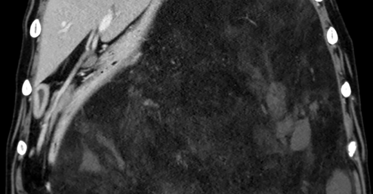
Miranda de la Vega, Mitch Robbins, Mark Howes, Miranda Vieson
Lipoma of the liver has not been reported in dogs. An 8 yr old spayed female Great Dane was referred for diagnostic workup of abdominal distention. Computed tomography showed fat-attenuating masses with negative attenuation values (variable between −60 to −40 Hounsfield units) and minimal contrast uptake within the left cranial abdomen. Left lateral and right medial liver lobectomies were performed to remove two liver masses. Histopathology showed large lipomas arising from within the hepatic parenchyma. Immunohistochemistry for smooth muscle actin was negative, consistent with true lipomas. The dog was euthanized 8 mo later because of causes likely unrelated to the liver lipoma. This is the first case report of lipoma in the liver of a dog. The purpose of this case report and brief literature review is to provide evidence that surgical excision of fat-attenuating masses within the liver that are consistent with lipoma using immunohistochemistry can be curative.
Rhonda Burge, Kevin D. Woolard, Jennifer L. Willcox, Robert B. Rebhun, Jenna H. Burton, Sami Al-Nadaf, Katherine A. Skorupski
Canine mast cell tumors (MCTs) have highly variable clinical behavior, and predicting outcomes in individual dogs remains challenging. Many studies combine dogs with varying tumor grades, clinical stage, or treatments, confounding those results. The purpose of this retrospective study was to determine outcome and prognostic factors in a specific subset of dogs with high-grade, stage 2, cutaneous MCTs treated with adequate local control via surgery with or without radiation therapy and adjuvant cytotoxic chemotherapy. Seventeen dogs met the inclusion criteria, and the median survival time was 259 days. Development of local recurrence, tumor location, and presence of ulceration were all associated with shorter survival times. Tumor size, mitotic count, chemotherapy protocol, lymph node classification, and radiation therapy were not significantly associated with outcome. In this study, a specific population of dogs characterized by high-grade MCTs with local lymph node metastasis who received aggressive local and systemic therapy had a median survival of about 8.5 mo. Dogs with ulcerated tumors, recurrent tumors, or tumors located on the head had a worse outcome despite aggressive therapy. These results may serve as a basis of comparison for future research exploring alternative treatment combinations in this specific population of dogs.
Tania Al Kafaji, Carlo Cantile, Fabio Tocco, Antonella Gallucci
An 11 yr old female French bulldog was presented for acute onset of seizures and a 2 wk history of disorientation. On physical examination, a nodular mass at the fourth mammary gland level was observed. Neurological evaluation showed obtundation and compulsive behavior. Brain MRI study did not reveal any abnormalities. Cerebrospinal fluid (CSF) collected from the cerebellomedullary cistern showed a marked increase of total nucleated cell count (400 cells/µL). Cytological evaluation identified the presence of a monomorphic round cell population characterized by large cell bodies, a single eccentrical located nucleus with high nuclear:cytoplasmatic ratio, and marked atypia with anisocytosis, anisokaryosis, and multiple nucleoli. Leptomeningeal carcinomatosis (LC) was suspected. The dog was euthanatized for worsening of clinical signs. Post-mortem examination identified an anaplastic mammary carcinoma in the nodular mammary mass. Infiltration by neoplastic cells exhibiting the same morphological features was detected along leptomeninges of the telencephalon and cerebellum associated with cortical and subcortical parenchymal micrometastases. To our knowledge, this is the first case of LC in a dog detected by CSF evaluation but without any MRI abnormalities. This finding emphasizes the usefulness of CSF cytology in patients with suspected LC even in the absence of any MRI identifiable lesions.
Andrea Nichole Mastorakis, Barbro Filliquist
A 1 yr old, 1.7 kg, spayed female Chihuahua was presented for respiratory distress and an enlarged cardiac silhouette as seen on thoracic radiographs. Echocardiogram revealed pericardial effusion and cardiac tamponade. Computed tomography revealed marked pleural and pericardial effusion, thickening of the pericardium caudally, and a mass along the mediastinum. Pericardial fluid obtained via pericardiocentesis showed suppurative inflammation with mixed anaerobic bacteria isolated on culture. Subtotal pericardiectomy and partial lung lobectomy was performed to treat septic pericarditis. Postoperative echocardiogram showed increased right-sided pressures consistent with constrictive epicarditis, and 10 days after surgery, the dog was re-presented for right-sided heart failure. An epicardectomy was performed. A definitive source of infection was not identified, although a penetrating foreign body (e.g., grass awn) was suspected. The dog recovered, and 10 yr follow up revealed no evidence of constrictive pathology on echocardiogram. This case report demonstrates the successful treatment of septic pericarditis and constrictive epicarditis via subtotal pericardiectomy and epicardiectomy.
Justin Mergl, Laura Nutt, Augusto Pareja
Two cats were presented with acute left-sided paresis after implantation of a microchip at the referring veterinary clinic. Neurological examinations were consistent with left-sided lesions between spinal cord segments C1 and C5. Orthogonal radiographs of the cervical spine showed a microchip dorsoventrally oriented, partially embedded in the cervical vertebral canal. Fluoroscopy was used to localize and retrieve the foreign body from the cervical spinal cord in each case. Improvement in clinical condition and return to ambulation was observed in both cats within 48 hr of surgical removal of the implant. No significant perioperative adverse events were noted during the surgical retrieval of the microchip. Two previously reported cases of intraspinal canal microchip placement had been treated surgically by hemilaminectomy. This approach carries a risk of complications, including hemorrhage from the venous sinus, iatrogenic damage to the spinal cord, and improper identification of the surgical site, requires advanced surgical training, and typically has an extended surgical time. The use of fluoroscopy to assist intraoperative localization of a spinal canal foreign body may lessen the requirement for more invasive surgical procedures.