
See the Full Issue
May 2023
Jan Bellows, DVM, DAVDC, DABVP and his team had to get creative when trying to intubate a dog with such severe periodontal disease that its mouth could not be opened far enough to see the larynx.
See the Full Issue
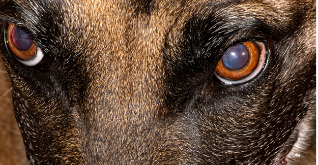
Figure 1: Swelling is evident beneath Tella’s left eye.
Tella, a seven-year-old, 21 kg rescue Belgian Malinois, presented for evaluation of a three-month intermittent swelling underneath her left eye (Figure 1). Oral administration of Clavamox prescribed by the referring veterinarian would temporarily resolve the swelling only to return once medication was discontinued. Tella’s other problem was that her mouth would not open greater than 15 mm. Her owners suspected previous trauma earlier in life before she was adopted (Figure 2).
Marked oral malodor was noted. Facial examination confirmed a broad soft swelling below the left eye. Fine-needle aspiration of the facial swelling revealed nondegenerative neutrophils, few macrophages, and cocci bacteria consistent with neutrophilic inflammation. Periapical periodontitis affecting the left maxillary fourth premolar was suspected. Intraoral buccal examination of the left upper cheek teeth revealed swelling, inflammation, and gingival recession of the surfaces surrounding the mesial palatal root of the left maxillary fourth premolar. (Figures 3a, 3b).
Ideally, the next step would have been to anesthetize for an in-depth, tooth-by-tooth examination including probing and intraoral radiographs. Unfortunately, this case presented two challenges: (1) how to intubate a dog whose mouth cannot be opened sufficiently to visualize the larynx; and (2) how to extract the maxillary fourth premolar if the mouth could not be opened sufficiently to access the tooth palatally.
The plan for anesthesia included tracheal intubation. If tracheal intubation was not possible, temporary tracheostomy was considered a secondary option. Before the clinical examination, Tella received 200 mg gabapentin, 50 mg trazadone, and Cerenia 1mg/kg. Butorphanol 0.2mg/kg was administered for premedication; methadone 0.1 mg/kg, midazolam 0.2mg/kg, and alfaxan 3.0mg/kg for induction. Isoflurane was used to maintain anesthesia.
Figure 2 (Left): Tella’s mouth could not be opened more than 15 mm.
Figure 3a (Middle): Clinical appearance of the left cheek teeth.
Figure 3b (Right): Swelling surrounding the mesiopalatal root of the left maxillary fourth premolar.
Figure 4a (Left): CO2 sampling tube placed over urinary catheter.
Figure 4b (Middle): CO2 reading consistent with endotracheal intubation.
Figure 4c (Right): Endotracheal tube threaded over the urinary catheter.
Figure 5a (Left): Confirmed advanced periodontal disease.
Figure 5b (Middle): CBCT imaging of the left temporomandibular joint area.
Figure 6a (Right): 701 surgical bur used to remove the crown of the mandibular first molar.
Figure 6b (Left): Hemisecting mandibular first molar before extraction.
Figure 7 (Middle): Postoperative CBCT imaging confirmed complete extractions.
Figure 8 (Right): Normal postoperative appearance of the left cheek teeth.
Tracheal intubation was initially accomplished by threading a #10 French plastic urinary catheter into the trachea with the neck extended. A normal carbon dioxide level (versus zero if esophageal intubation) confirmed tracheal location of the catheter (Figures 4a, 4b, 4c). An endotracheal tube was then threaded over the urinary catheter.
Once tracheal intubation was confirmed, cone beam computed tomography (CBCT) imaging was performed, which showed advanced periodontal and periapical disease of the left maxillary fourth premolar, advanced periodontal disease of the second molar (which was virtually floating in the caudal oral cavity), as well as the left mandibular first, second, and third molars (Figure 5a). Further CBCT evaluation revealed a large callus just rostral to the left temporomandibular joint, which explained why Tella could only partially open her mouth (Figure 5b).
The left maxillary fourth premolar, second molar, and the mandibular first, second, and third molars were extracted, approaching all teeth buccally. To aid the extraction process, the crowns were removed, then the roots (Figures 6a, 6b). Postoperative CBCT imaging was performed confirming complete extractions. (Figure 7).
Tella was sent home on gabapentin and meloxicacm. No antimicrobials were prescribed. On re-examination two weeks postoperatively, the swelling below the left eye had resolved, and all surgical sites appeared to be healed. Tella’s owners are considering surgery to remove a section of the left caudal mandible to allow her to facilitate a larger mouth opening and future dental procedures to address the remaining teeth affected by moderate to advanced periodontal disease (Figure 8).
Jan Bellows, DVM, DAVDC, DABVP, is owner of AAHA-accredited Hometown Animal Hospital and All Pets Dental Clinic in Weston, Florida. |
Photo credits: Photos courtesy of Jan Bellows, DVM, DAVDC, DABVP