
See the Full Issue
September 2023
Abstracts from the latest issue of the Journal of the American Animal Hospital Association.
See the Full Issue
Monique Triglia, Hollie Horton, Melanie Dobromylskyj, Amy Jo Ferreira, William Peter Robinson
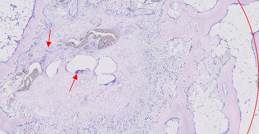
A 4 yr old female neutered Labrador retriever was referred with a history of left hind-limb lameness and an acute, nonpainful, subcutaneous mass on the medial aspect of the left stifle. Stifle radiographs and fine-needle aspirates of the soft-tissue mass performed by the referring veterinarian confirmed the presence of predominantly highly granulated mast cells, consistent with a mast cell tumor. Computed tomography demonstrated a soft-tissue mass centered on the left medial stifle, with associated joint effusion and polyostotic lytic lesions on the tibial plateau and distal patella. Ultrasound-guided aspirates of the liver, spleen, and popliteal lymph nodes were obtained to rule out further metastatic spread. Cytology of the joint fluid demonstrated a low number of well-differentiated mast cells. Surgical and oncological interventions were discussed, and full hind-limb amputation was elected. Histopathological analysis of the submitted tissues after amputation diagnosed a subcutaneous mast cell tumor with neoplastic cell infiltrate extending into sections of joint capsule and synovial membrane. Infiltration to the tibia and distal patella were suspected following the presence of mast cell clusters in both osteolytic lesions. No evidence of metastasis was identified in the popliteal lymph node. Postoperative monitoring of iliac lymph node size using ultrasound did not identify evidence of metastasis 12 mo postoperatively.
Karen Marina Hernandez Guzman, Kenneth Harkin
Acquired myasthenia gravis (MG) in dogs can present with focal or generalized weakness and is diagnosed by the presence of circulating antibodies to the acetylcholine receptor. Megaesophagus is the most common focal form of MG. Although exacerbation of MG has been associated with the use of fluoroquinolones in humans, it has not been previously described in dogs. The medical records of 46 dogs diagnosed with MG based on acetylcholine receptor antibody testing from 1997 to 2021 were retrospectively evaluated to identify any dogs who demonstrated exacerbation of MG after the administration of a fluoroquinolone. Exacerbation of MG, from focal to generalized, occurred in a median of 4.5 days after initiation of fluoroquinolone therapy in six dogs. In addition, one dog with generalized MG and megaesophagus developed pyridostigmine resistance subsequent to fluoroquinolone therapy. Marked improvement in generalized weakness was reported 36 hr after discontinuation of fluoroquinolone therapy alone in one dog and in combination with pyridostigmine in two dogs. Fluoroquinolone therapy was never stopped in three dogs who were euthanized because of severe weakness and one dog who died of respiratory arrest.
Marion Signoret, Clément Musso, Harriet Hahn, Kévin Le Boedec, Laurent Cauzinille
Accurate diagnostic imaging is required for surgical planning of acute thoracolumbar intervertebral disc extrusion. However, data comparing the accuracy of conventional IV contrast-injected computed tomography (CT) and myelography CT for hemilaminectomy localization and size assessment are sparse. In this study, IV contrast-injected CT and myelography CT were performed in 48 French bulldogs presenting with acute disc herniation and compared with postsurgical CT. CT images were evaluated by three raters. IV contrast-injected CT erroneously identified the compressive lesion site significantly more often than myelography CT. The length of the compressive lesion was significantly higher using myelography CT compared with conventional CT, but this did not lead to relevant consequences on the surgical opening site length. Myelography CT should therefore be recommended when thoracolumbar disc disease is suspected and multiple compressive lesions are visualized on IV-injected CT in French bulldogs.
Jennifer Clegg, Clarissa Souza, Bailey Brame
Otitis externa (OE) is a common disease in dogs, and topical medications are the preferred treatment. Compounded solutions of enrofloxacin are commonly used in practice to treat bacterial OE; however, the tolerability of different concentrations of this antibiotic in the ear canals of dogs has not been evaluated. The objective of this study is to determine if a higher concentration of enrofloxacin applied to the external ear canal is clinically tolerated in dogs with healthy ears. Sixteen client-owned dogs with bilateral healthy ears and no previous history of OE were enrolled. Injectable enrofloxacin 2.27% diluted with sterile sodium chloride in 1:1 (11.35 mg/mL) and 2:1 (15 mg/mL) ratios were applied into the dogs’ right and left ears, respectively, q 12 hr for 14 days. Based on video otoscopic examination, clinical score for canine OE (OTIS3) results before application were ≤1 for all dogs. During the study and at the conclusion, all scores remained ≤2, which is considered normal. No cytologic inflammatory cells were seen in any of the dogs’ ears throughout the study. Different concentrations of enrofloxacin solution applied topically were well tolerated by dogs with healthy ears and can be considered for the treatment of dogs with bacterial OE.
Christen Montesanto, Brenda Phillips, Oceane Aubry
A 2 yr old female intact flat-coated retriever dog was presented for evaluation of a histologically diagnosed cutaneous Langerhans cell histiocytosis of the muzzle with right mandibular nodal metastasis and suspected prescapular lymph node metastasis. Chemotherapy (lomustine 60 mg/m2 by mouth as a single dose) and glucocorticoid therapy (prednisone ~20 mg/m2 by mouth every 24 hr) were initiated. Progressive disease occurred 21 days after lomustine administration. Doxorubicin (at 30 mg/m2 IV every 3 wk) was administered as a second-line therapy. Prednisone was continued at the same dose. Partial response was noted 1 wk after initiation of doxorubicin and sustained through doxorubicin #2. Complete remission was achieved following doxorubicin #3 (63 days from the start of doxorubicin rescue therapy). Progressive disease was noted after doxorubicin #5, for a total duration of response to doxorubicin of 105 days. Further rescue treatment with vinorelbine at 15 mg/m2 IV was elected. Progressive disease and clinical decline were noted 1 wk after initiation of vinorelbine. The patient was euthanized because of clinical decline 126 days after histopathologic diagnosis and 114 days after chemotherapy treatment was initiated.
Michael Jaffe, Kayla Alexander, Michelle Ryan, Blair Bennett, Brittany Baughman
A 4 mo old male goldendoodle puppy was evaluated for chronic hematochezia with a history of recurrent rectal prolapse and tenesmus. A colo-colonic intussusception was diagnosed via abdominal imaging. Surgery was elected to reduce the intussusception, wherein a colonic mass was discovered. Colonic resection and anastomosis was performed, and the tissues were submitted for histopathological examination. The puppy was diagnosed with colonic hamartomatous ganglioneuromatosis based on the presence of markedly hyperplastic submucosal and myenteric plexi with infiltration and expansion of the mucosa and submucosa by Schwann cells and neuronal cell bodies. Ganglioneuromatosis is a rarely reported entity in the veterinary literature, and limited clinical follow-up data is available for described cases. In humans, ganglioneuromatosis is associated with a PTEN genetic mutation, which confers increased susceptibility to the development of neoplasia of endocrine organs. Approximately 1 yr after the operation, this puppy appeared clinically normal with no abnormalities on repeated imaging. This case report describes the clinical presentation, surgical treatment, and histologic features of colonic hamartomatous ganglioneuromatosis with 1 yr postoperative clinical follow up data in a dog. Although uncommon, ganglioneuromatosis should be considered as a differential diagnosis list as a cause of gastrointestinal masses in puppies and young dogs.
Mengxuan Zhang, Penny J. Regier, Jackson Sanders
A 1 yr old castrated male shih tzu was referred for recurrent urinary tract infections (UTI), prostatitis, and urine dribbling that was not responsive to medical management. Physical examination and computed tomography scan revealed a micropenis with a disproportionately high prepuce-to-penis ratio. Preputial shortening with a hexagonal, full thickness preputial resection followed by preputial anastomosis was performed. The dog recovered from surgery with no complications. Urine dribbling persisted in the short-term postoperative period, but the patient achieved significant clinical improvement and resolution of his urine dribbling and recurrent UTIs at the 1 yr follow-up. In conclusion, this surgical technique was able to successfully restore quality of life in a dog with a micropenis, and preputial shortening should be considered in cases of recurrent UTIs where there is significant disparity between the size of the penis and the prepuce.
Joonseok Kim, Yeon Chae, Dohee Lee, Yoonhoi Koo, Sijin Cha, Taesik Yun, Mhan-Pyo Yang, Byeong-Teck Kang, Hakhyun Kim
It has been reported that hypertriglyceridemia can partially mediate between diabetes mellitus (DM) and pancreatitis in dogs, implying that another mediator, such as chronic hyperglycemia, might exist. Therefore, this study aimed to evaluate the relationship between hyperglycemia and serum canine pancreatic lipase immunoreactivity (cPLI) concentration in diabetic dogs. This retrospective cohort study included 26 client-owned diabetic dogs, divided according to their serum fructosamine levels (<500 μmol/L = well-controlled DM group; ≥500 μmol/L = untreated or poorly controlled DM group). Five of the 26 DM dogs (19.2%) had serum cPLI concentrations consistent with pancreatitis, among which two showed ultrasonographic evidence of pancreatitis without clinical signs. The serum cPLI concentrations (median [interquartile range]) were significantly higher in the untreated or poorly controlled group (520 μg/L [179.76–1000 μg/L]) than in the well-controlled group (77 μg/L [32.22–244.6 μg/L], P = 0.0147). The serum fructosamine concentration was positively correlated with the serum cPLI concentration (r = 0.4816; P = 0.0127). Multivariate analysis revealed serum triglyceride and fructosamine concentrations were associated with the serum cPLI concentration. In conclusion, this study suggests that chronic hyperglycemia may induce pancreatic inflammation in diabetic dogs; however, the clinical significance of increased cPLI concentration is unknown.